
 Volume 112, Current Issue
 Vol 28, October 2019
 Vol 27, September 2019
 Vol 26, August 2019
 Vol 25, July 2019
 Vol 24, June 2019
 Vol 23, May 2019
 Vol 22, April 2019
 Vol 21, March 2019
 Vol 20, February 2019
 Vol 19, January 2019
 Vol 18, December 2018
 Vol 17, November 2018
 Vol 16, October 2018
 Vol 15, September 2018
 Vol 14, August 2018
 Vol 13, July 2018
 Vol 12, June 2018
 Vol 11, May 2018
 Vol 10, April 2018
 Vol 9, March 2018
 Vol 8, February 2018
 Vol 7, January 2018
 Vol 6 No 12, December 2017
 Vol 6 No 11, November 2017
 Vol 6 No 10, October 2017
 Vol 6 No 9, September 2017
 Vol 6 No 8, August 2017
 Vol 6 No 7, July 2017
 Vol 6 No 6, June 2017
 Vol 6 No 5, May 2017
 Vol 6 No 4, April 2017
 Vol 6 No 3, March 2017
 Vol 6 No 2, February 2017
 Vol 6 No 1, January 2017
 Vol 5 No 12, December 2016
 Vol 5 No 11, November 2016
 Vol 5 No 10, October 2016
 Vol 5 No 9, September 2016
 Vol 5 No 8, August 2016
 Vol 5 No 7, July 2016
 Vol 5 No 6, June 2016
 Vol 5 No 5, May 2016
 Vol 5 No 4, April 2016
 Vol 5 No 3, March 2016
 Vol 5 No 2, February 2016
 Vol 5 No 1, January 2016
 Vol 4 No 12, December 2015
 Vol 4 No 11, November 2015
 Vol 4 No 10, October 2015
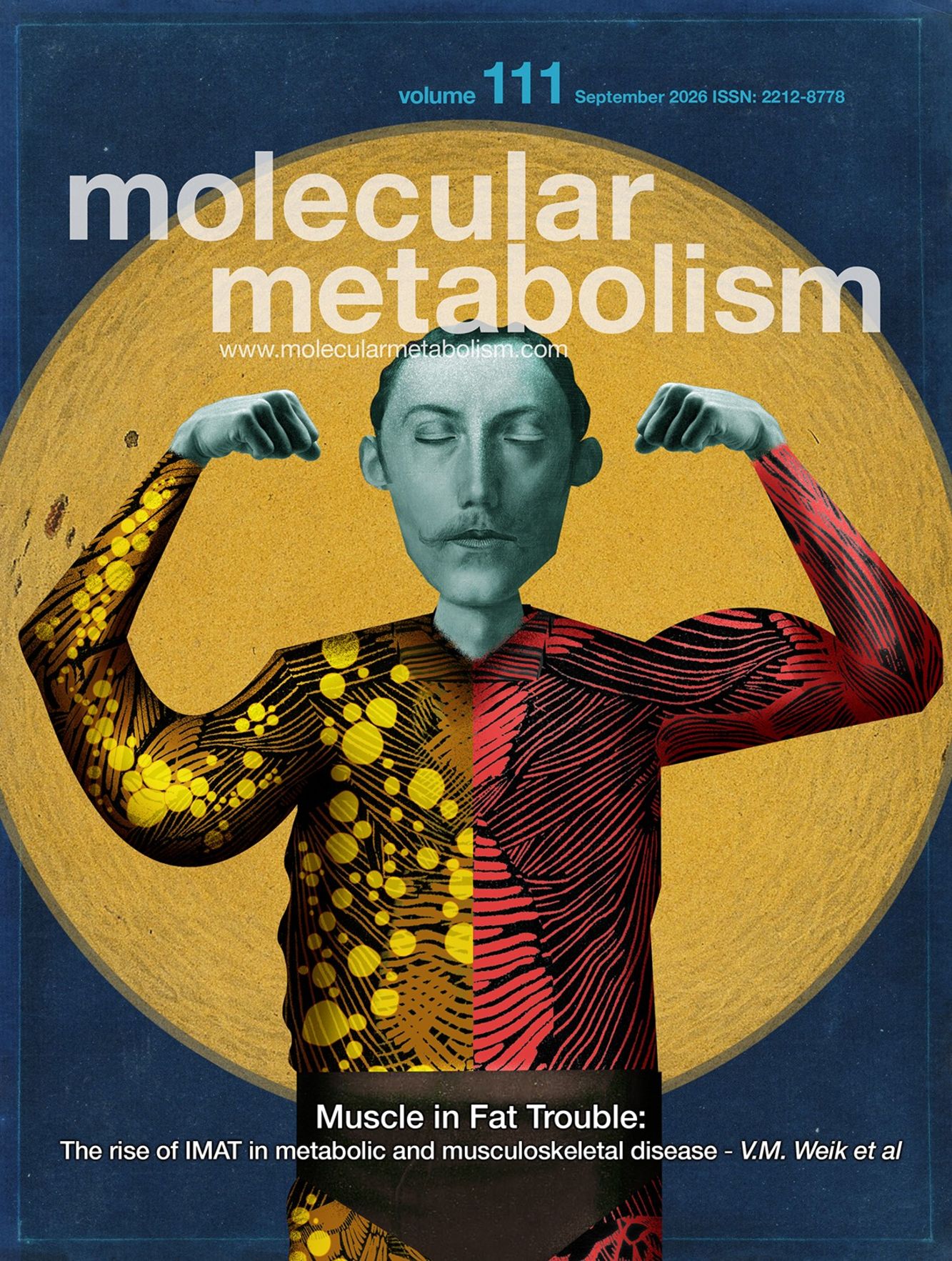
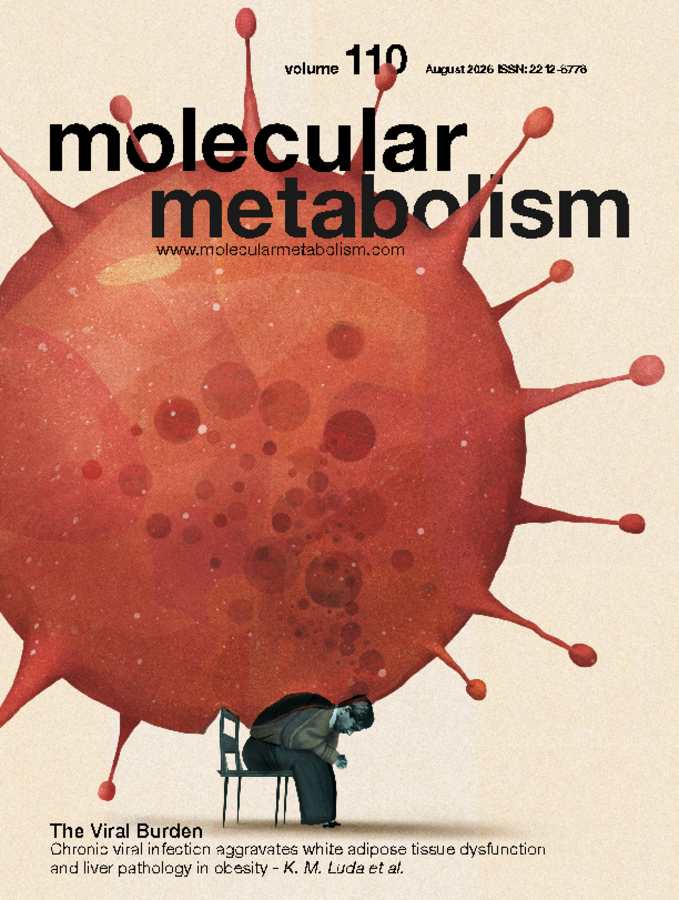

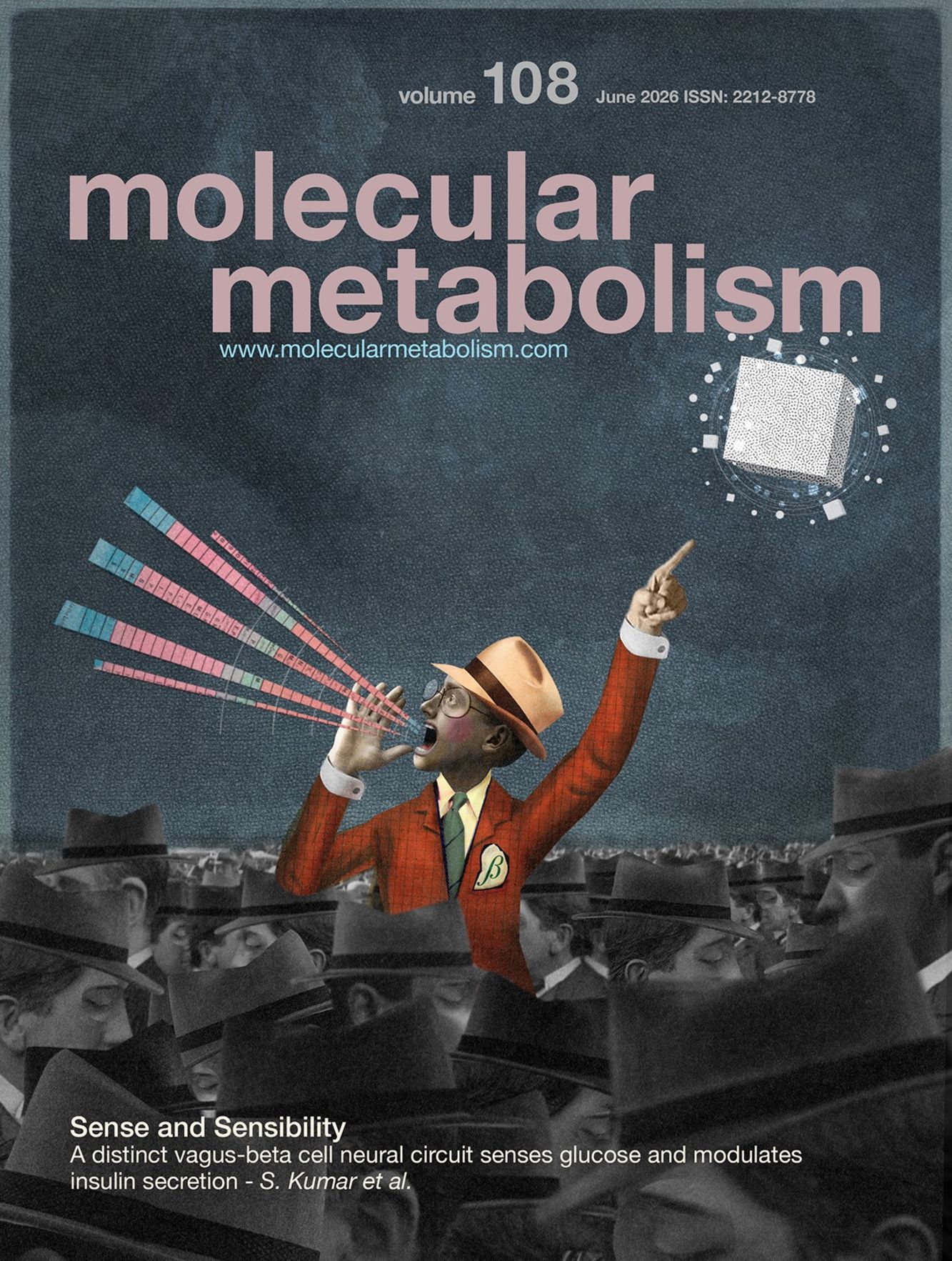

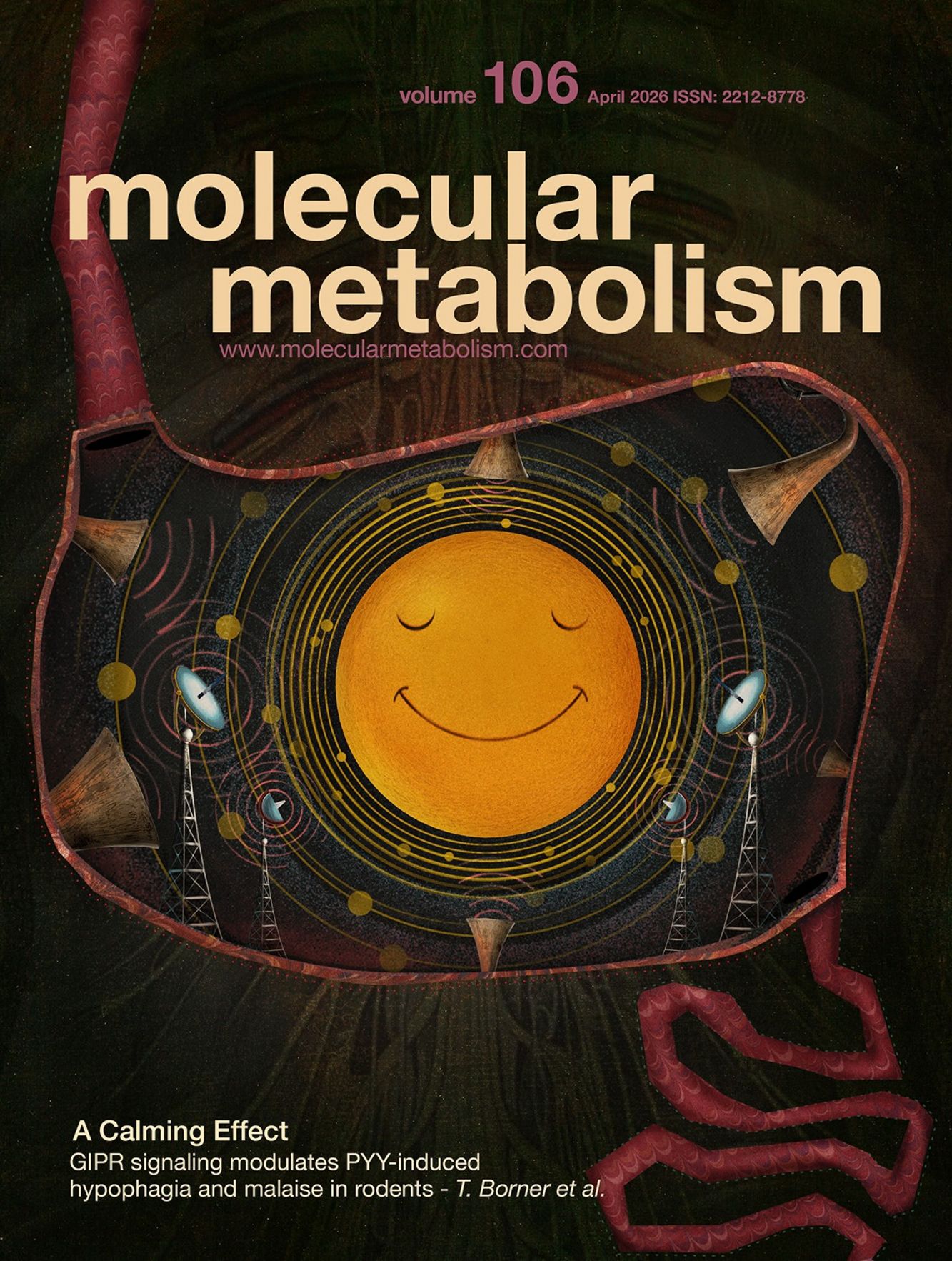












Cover Story Current Issue
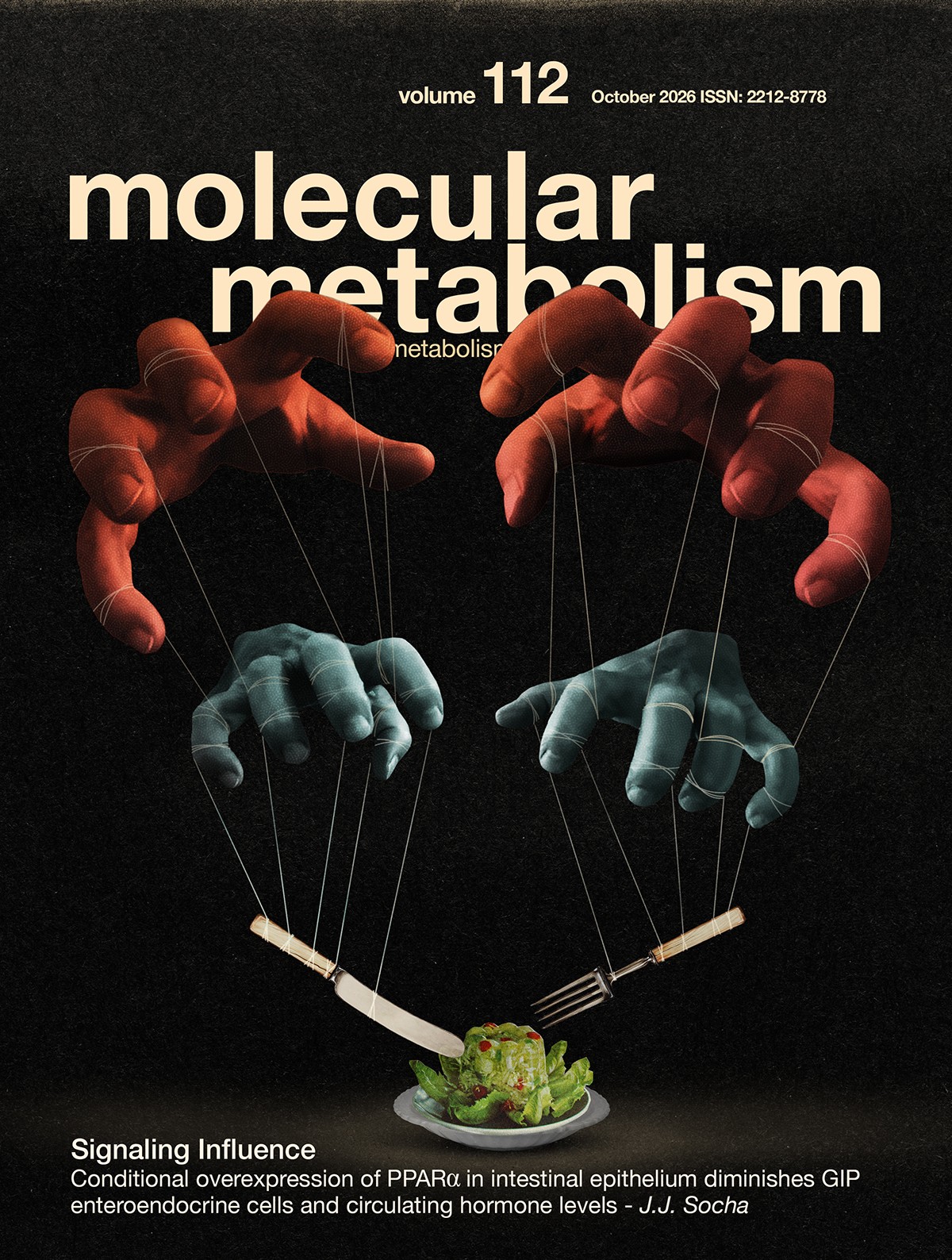
The small intestine, as the primary site of nutrient absorption, integrates signals from dietary components and gut microbiota to coordinate systemic energy balance, metabolism, and glucose homeostasis alongside other tissues such as the pancreas, liver, and brain. Nuclear receptors represent one mechanism by which the intestinal epithelium senses and respond to dietary signals via transcriptional regulation of metabolic programs. The Peroxisome Proliferator Activated Receptors (PPARs), including PPARα, PPARδ, and PPARγ, are lipid-responsive nuclear hormone receptors. PPAR transcriptional activity is highly context-dependent, shaped by the availability and affinity of their lipid ligands and co-regulators as well as by cell and tissue types. Whereas PPARγ and PPARδ are established regulators of glucose homeostasis, acting in white adipose tissue, skeletal muscle, and liver to improve insulin sensitivity, PPARα has been primarily associated with hepatic lipid metabolism, where it promotes fatty acid transport, β-oxidation, and ketogenesis. Due to their roles in lipid metabolism and anti-inflammatory processes, selective PPAR agonists are being actively pursued as therapies in clinical and pre-clinical studies for metabolic diseases. Thus, it is critical to improve our understanding of the context-specific and tissue-specific effects of PPAR signaling.
Current Issue
- Abstract
Adaptive oligodendrogenesis regulates blood-hypothalamus barrier permeability, hypothalamic glucose sensing and systemic glucose homeostasis in male mice
Objective
Brain glucose sensing is critical for survival during hypoglycaemia, yet how glucose-sensing neurons access circulating glucose concentrations to maintain glucose homeostasis remains poorly understood. Here we tested the hypothesis that adult oligodendrogenesis in the median eminence (ME) is responsive to changes in blood glucose levels and contributes to hypothalamic glucose sensing through regulation of the blood-hypothalamus barrier.
Methods
We used glycemic challenges and hypoinsulinaemic clamp studies to identify the effect of systemic changes in glycaemia on hypothalamic oligodendrocyte lineage cells. We used conditional knockout mouse models to dissect the respective contributions of adult oligodendrogenesis and new myelin formation to glucose homeostasis in adult male mice. Analyses combined immunofluorescence, serial electron microscopy, whole-brain tissue clearing, and transcriptomic analyses.
Results
We found that adult oligodendrogenesis in the median eminence (ME) is modulated by changes in circulating glucose levels and rapidly upregulated by hypoglycaemia. Genetic blockade of new oligodendrocyte production in adult male mice impairs the regulation of glucose homeostasis, the integrity of the ME blood-hypothalamus barrier, and hypothalamic glucose sensing. Unexpectedly, functional integrity of adult-formed myelin is not required for the maintenance of glucose homeostasis. Instead, we show that blockade of adult oligodendrogenesis disrupts hypothalamic expression of A disintegrin and metallopeptidase with thrombospondin motifs 4 (ADAMTS4), a metallopeptidase whose brain expression is restricted to the oligodendrocyte lineage and whose ME expression requires ongoing adult oligodendrogenesis. We show that ADAMTS4 regulates hypothalamic perineuronal net deposition, vascular permeability and glucose sensing. Finally, we show that ME ADAMTS4 expression is regulated by changes in peripheral glycaemia and is dysregulated in diabetes, providing a mechanism by which ME oligodendrocytes contribute to the regulation of glucose homeostasis.
- Abstract
Conditional overexpression of PPARα in intestinal epithelium diminishes GIP enteroendocrine cells and circulating hormone levels
Agonists of the lipid-sensing PPAR nuclear receptors, including PPARα, are being explored as therapies for metabolic disorders due to their roles in metabolic and anti-inflammatory processes. PPARα transcriptional programs have tissue-dependent features, yet there is a lack of genetic tools to study tissue-specific signaling activation without the addition of a systemic agonist. We aimed to investigate intestinal epithelial cell (IEC)-specific roles of PPARα signaling using a novel transgenic mouse that enables spatial and temporal control of Ppara overexpression. CAG-Ppara,-EGFP mice were bred to Villin-CreERT2 to establish the IEC-Ppara mouse, which was compared to littermate controls 2 weeks after tamoxifen exposure. IEC-Ppara mice had increased Ppara mRNA and PPARα protein in the intestinal epithelium. Transcriptional analysis of intestinal tissue from IEC-Ppara mice showed upregulation of PPARα target genes and functional enrichment for fatty acid catabolic processes. As expected, the enterocytes of IEC-Ppara mice were primed to absorb lipids following oral administration of an olive oil bolus. Unexpectedly, the enteroendocrine hormone Gip was among the most downregulated genes. GIP-positive cells were reduced in the intestines of IEC-Ppara mice and in mice treated with PPARα agonist WY-14643. Circulating GIP hormone was reduced in IEC-Ppara mice. GLP-1-positive cells and hormone were unchanged. Consistent with reduced GIP function, IEC-Ppara mice consumed more food. These findings reveal PPARα as a regulator of GIP and support a new framework in which PPARα signaling influences systemic energy balance via a gut hormone axis. This study could have future impact on understanding responses to therapies targeting PPAR or incretin signaling.
- Abstract
Targeting microbial bile salt hydrolase reprograms bile acid metabolism and ameliorates metabolic dysfunction–associated steatohepatitis in mice
Microbial bile salt hydrolase (BSH) plays a central role in shaping bile acid composition and gut–liver metabolic signaling, yet its therapeutic potential in metabolic dysfunction–associated steatohepatitis (MASH) remains incompletely defined. Here, we evaluated the efficacy of the non-absorbable BSH inhibitor GR-7 in a diet-induced mouse model of steatohepatitis using early and late intervention strategies with different dosing regimens. GR-7 reduced food intake and exerted stage- and dose-dependent therapeutic effects, with early intervention robustly suppressing hepatic fibrosis even at a low dose, whereas late-stage administration of high-dose GR-7 markedly reduced hepatic steatosis and inflammation, as evidenced by decreased liver weight, hepatic triglyceride and cholesterol levels, and plasma ALT. Although late intervention did not result in statistically significant histological reversal of fibrosis, a trend toward improvement was observed, together with suppression of fibrogenic gene expression, suggesting that prolonged treatment may further enhance antifibrotic efficacy. Mechanistically, GR-7 effectively inhibited microbial BSH activity in vivo, leading to reduced cecal unconjugated primary and secondary bile acids—including deoxycholic acid and lithocholic acid, which was associated with improved gut barrier integrity and reduced hepatic inflammation. In parallel, BSH inhibition reprogrammed hepatic bile acid metabolism toward activation of the alternative CYP27A1-mediated synthesis pathway, accompanied by reduced food intake, thereby contributing to reduced hepatic lipid accumulation. Furthermore, late-stage high-dose treatment selectively remodeled the hepatic immune landscape rather than fully restoring homeostasis, highlighting immune recalibration as a key component of therapeutic response. Together, these findings identify microbial BSH inhibition as a promising microbiome-targeted therapeutic strategy for MASH.
- Abstract
Human brown fat metabolism associates with systemic branched-chain amino acids homeostasis
Circulating branched-chain amino acids (BCAAs) are linked with insulin resistance, but the human tissues contributing to systemic BCAA homeostasis remain incompletely defined. Brown adipose tissue (BAT) is a metabolically active adipose depot associated with favourable insulin sensitivity, yet its role in BCAA metabolism in humans remains unclear. We tested whether human BAT metabolism is associated with circulating BCAA levels, BAT-resident BCAA-catabolic signatures, and longitudinal changes in systemic BCAA homeostasis. We studied 83 adults who underwent metabolic phenotyping, PET-CT assessment of cold-stimulated BAT metabolism, and serum metabolomic profiling at room temperature and during acute mild cold exposure. Supraclavicular BAT biopsies from 25 participants were analysed by transcriptomics and metabolomics, and 40 participants were re-examined for circulating BCAA profiles after approximately five years. Participants with high BAT metabolism had lower circulating BCAA levels than those with low BAT metabolism. Within BAT, metabolically active individuals exhibited lower relative BCAA abundance together with higher expression of genes involved in BCAA catabolism. These BAT BCAA-catabolic signatures aligned with thermogenic capacity and indices of systemic insulin sensitivity. In contrast, individuals with low BAT metabolism showed increases in circulating BCAAs over five years. Integrative analyses further linked circulating lipopolysaccharide, a marker of metabolic endotoxemia, with higher BAT BCAA and aminomalonate abundance, together with transcriptional patterns involving inflammatory and mitochondrial pathways. Together, these findings identify human BAT metabolism as a tissue phenotype linked to systemic BCAA homeostasis and extend the role of human BAT beyond thermogenesis, suggesting that BAT-associated BCAA handling may contribute to systemic metabolic health.
Articles in Press
- Abstract
Triple-negative breast cancer (TNBC) is a highly aggressive breast cancer subtype with limited targeted therapies, leading to higher mortality compared with other breast cancer subtypes. In our previous study we identified N-acyl sphingosine amidohydrolase 1 (ASAH1) to be overexpressed in TNBC cells and demonstrated its role in promoting TNBC growth and progression. ASAH1 is a metabolic enzyme that converts ceramide into sphingosine and free fatty acids, thereby creating a favorable environment for tumor growth. To gain a more comprehensive understanding of the metabolic alterations associated with ASAH1 inhibition, we performed a large-scale metabolomic analysis of TNBC cells expressing ASAH1 shRNAs. Our analysis revealed a significant increase in carnosine levels following ASAH1 inhibition. Subsequent studies demonstrated that exogenous treatment of TNBC cells with carnosine inhibited their growth. Moreover, combining carnosine with ASAH1 inhibitors (carmofur or ceranib-2) resulted in potent synergistic inhibition of TNBC growth. Mechanistically, the combined treatment led to a greater reduction in mitochondrial membrane potential, increased mitochondrial superoxide production and further increased apoptosis. Collectively, these results identify a new metabolism-based combination therapeutic strategy for the effective treatment of TNBC.
- Abstract
To investigate the link between skin features and diabetes, we performed a multi-modal study combining a diabetic mouse model, a human pilot cohort, and keratinocyte response assays. Our goal was to identify molecular signatures in skin, cells, and sweat that correlate with glycemic control and could serve as early biomarkers of hyperglycemia. This integrative approach advances understanding of diabetes-associated dermatological changes and supports the development of targeted, non-invasive diagnostic tools for early detection.
Following glucose tolerance tests, proteomic and metabolomic profiling was performed. We identified dihydrolipoyl-succinyltransferase (DLST) as a promising biomarker whose protein and transcript levels consistently correlated with glycemic status across models. In HaCaT keratinocytes, DLST was found to drive metabolic reprogramming toward glutamine utilization as part of an antioxidant protective response. Chromatin immunoprecipitation assay revealed that Sp1 binds to the DLST promoter region, while functional studies, including Sp1 overexpression and knowdown, further confirmed that Sp1 regulates DLST at transcriptional level. Furthermore, DLST knockdown reproduced metabolic shifts in glutamine pathways, findings supported by proteomic analyses and stable isotopic flux analysis.
Reduced DLST expression in healthy skin corresponded with glutamine metabolism changes observed in keratinocytes upon oral glucose ingestion. While healthy skin and sweat displayed opposing metabolic trends, pre-type 2 diabetes samples showed aligned trends, suggesting sweat metabolomics could serve as an early systemic metabolic indicator.
These findings identify DLST as a glucose-responsive biomarker reflecting skin and sweat responses to systemic glucose, providing a foundation for the development of targeted, non-invasive diagnostic tools for early detection of diabetes.
- Abstract
Objective
Cholecystokinin (CCK) has been implicated in coordinating lipid digestion and satiety. However, the molecular mechanisms underlying nutrient-dependent CCK-release from human I-cells are not well understood, at least in part due to the cells’ scattered distribution and a lack of reliable CCK-assays. Here we used human intestinal organoids to characterize lipid-sensing mechanisms underlying CCK release.
Methods
Human duodenal organoids were genetically engineered using CRISPR-Cas9 to insert either the fluorescent protein Venus or the cAMP reporter Epac-S-H187 at the CCK locus. Transcriptomic profiling of CCK-positive and negative cells was performed following fluorescent-activated cell sorting. Intracellular calcium and cAMP as well as CCK-GRAB-sensor secretory responses were assessed during live-cell imaging, and CCK secretion was quantified by LC-MS/MS. To evaluate lipid-sensing pathways, FFAR1, FFAR4, and GPR119 knockout organoids were generated.
Results
Transcriptomic analysis identified expression of lipid-sensing GPCRs in I-cells, including FFAR1, FFAR4, FFAR2, GPR119, GPBAR1, OR51E1 and OR51E2. Fatty acids and FFAR1 agonists increased intracellular calcium, whereas agonists of GPBAR1 and GPR119, and short-chain fatty acids elevated cAMP. CCK release was triggered by FFAR1, GPBAR and GPR119 agonists, and fatty acids with chain-length > C8. FFAR1 knockout organoids exhibited impaired Ca2+ and CCK secretory responses to fatty acids, whereas responses of FFAR4 KO organoids were like wild-type.
Conclusions
In human I-cells, FFAR1 plays a crucial role in lipid-induced CCK release, whereas FFAR4 appears to be redundant in this context. These findings improve understanding of human CCK physiology and identify potential therapeutic targets for metabolic regulation and appetite control.
- Abstract
Type 2 Diabetes (T2D) is a complex disease that has increased in prevalence in the last years affecting over 530 million people around the world. T2D increases mortality and morbidity by different mechanisms. As a complex disease, it is caused by many genetic changes and, consequently, T2D genetics has been analyzed from different points of view and using different strategies. Most studies have focused on inherited Single Nucleotide Variants (SNVs) present in genomic DNA, while other types of genetic variants have received less attention.
The aim of the present review is to examine the limitations of the studies conducted to date, and the importance of non-SNVs and non-germinal variants in the genetics of this disease. In this way, structural variants, mobile elements and tandem repeats have a crucial role in T2D. In addition, variations in mitochondrial DNA and somatic variations have been shown to play an important role in metabolic alterations that may lead to the development of T2D.
- Abstract
Pancreatic beta cells have the unique function of synthesizing and secreting high amounts of the inhibitory neurotransmitter γ-aminobutyric acid (GABA). The mechanism of GABA secretion, whether vesicular or channel-mediated, is debated. Our study reveals surprising temporal complexity in the pattern of islet GABA secretion. We used insulin secretion modulators to demonstrate that GABA release is not directly correlated with insulin secretion. VGAT reporter mice also showed that beta cells do not express the requisite vesicular GABA transporter (VGAT) for vesicular GABA release. Instead, GABA is secreted from the cytosol in pulses that depend on expression of the LRRC8D subunit of the volume regulatory anion channel (VRAC). We further demonstrate the dynamic coordination of GABA release with calcium influx in beta cells and dependence on beta cell depolarization. These results suggest a model where GABA is released during the peaks of beta cell calcium oscillations to provide feedback which strengthens and reinforces the oscillation waveform.
- Abstract
Background
β-klotho (KLB), the obligate co-receptor for fibroblast growth factor (FGF) 19 and 21 signalling, has emerged as a therapeutic target for metabolic disease. Current understanding of KLB function is based largely on germline knockout mouse models, where developmental abnormalities obscure its function in adult physiology. Here, we used somatic hepatocyte-specific gene editing in adult mice to define hepatic KLB function.
Methods
Hepatocyte-specific KLB deficiency was induced by retro-orbital delivery of adeno-associated virus particles containing guide RNAs targeting Klb in adult male mice expressing Cas9 in the liver. Following transduction, mice were maintained on a 4-week chow diet or a 12-week high-fat diet, prior to comprehensive metabolic and biochemical phenotyping.
Results
Adult-onset hepatic KLB deficiency did not alter body weight or composition, indicating that reduced bodyweight in germline models reflects developmental effects. In line with previous studies, KLB deficiency resulted in derepressed bile acid synthesis, elevated faecal bile acid excretion, and a shift towards a more hydrophobic bile acid pool. Importantly, we also uncovered a role for hepatic KLB in regulating lipid homeostasis. Chow-fed KLB-deficient mice exhibited reduced circulating very-low-density lipoprotein levels (VLDL) with marked hepatic triglyceride accumulation, suggesting altered VLDL metabolism as a potential mechanism driving steatosis.
Conclusions
Using an adult-onset liver-specific knockout model, we disentangled KLB function from developmental confounders and established KLB as a critical regulator of adult lipid and lipoprotein homeostasis. Our study redefines the physiological role of KLB in hepatic metabolism and has important implications for the interpretation of FGF19/KLB-directed therapies for metabolic diseases.
- Abstract
The molecular circadian clock coordinates cellular and systemic metabolism across the day, yet how it integrates with energy-sensing pathways in human skeletal muscle remains incompletely understood. AMP-activated protein kinase (AMPK) functions as a cellular energy sensor and has been implicated in circadian regulation; however, its role in coupling clock components to metabolic programming in human muscle has not been established.
Here, we investigated the interaction between AMPK signaling and the circadian machinery in primary human myotubes, focusing on the downstream clock components NPAS2 and DBP. Silencing of the AMPKα subunit altered core clock gene expression, increasing CRY1 and CRY2 while reducing NPAS2 and DBP expression, with rhythmicity preserved. To determine the functional relevance of these changes, we examined metabolic phenotypes following siRNA-mediated silencing of NPAS2. NPAS2 knockdown reduced basal oxidative metabolism, decreased abundance of electron transport chain subunits, and increased β-oxidation flux, while maximal mitochondrial respiratory capacity remained intact. These alterations occurred independently of canonical AMPK–ACC signaling but were associated with reduced HIF1A expression. In contrast, DBP silencing reduced glucose oxidation but did not recapitulate the broader metabolic remodeling observed with NPAS2 knockdown.
Together, these findings identify NPAS2 as a downstream mediator linking AMPK-dependent circadian signaling to skeletal muscle metabolic regulation. Disruption of this axis alters substrate utilization and impairs mitochondrial function, revealing a mechanism by which circadian clock components contribute to metabolic regulation in human skeletal muscle.

2025 Impact Factor: 6.3
You are what you eat
Here is a video of Vimeo. When the iframes is activated, a connection to Vimeo is established and, if necessary, cookies from Vimeo are also used. For further information on cookies policy click here.